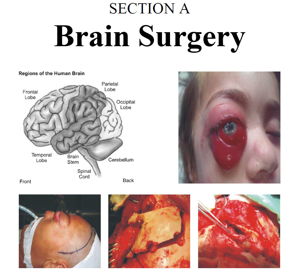
Navigating Brain Lesions Via Craniometric Points of Human Skull. Effectiveness in Targeted Craniotomy
DOI:
https://doi.org/10.36552/pjns.v24i1.417Keywords:
Craniotomy, Craniometric points, Human SkullAbstract
Background and Objectives: Navigating brain pathologies via external landmarks helps in guiding neurosurgeon to perform and plan brain surgeries without feeling compromised in their competence, when modern neuronavigation is not available. We assessed the navigating art of human craniometry in brain surgery, in terms of accuracy and safety, of our patients.
Materials and Methods: A descriptive study was in Department of Neurosurgery Quaid-e-Azam International Hospital on utilizing the art of Craniometry in navigating brain lesions and to see how effective this art is in the execution of neurosurgical procedures.
Results: Total of hundred patients were subjected in our study, who were according to inclusion criteria. Mean age calculated was 44 years with STD ± 6, with minimum age of 8months and maximum age of 83years. There were 61% male, and 39% female patients. Supratentorial lesions were 72%, ventriculoperitoneal shunts 11%,
and 17% were infratentorial.In 89 surgeries, we were exactly on the target according to our external skull landmarks, in 4 cases we slightly deviated about 0.3 cm, and in 7 surgeries we encounter some difficulty in navigating lesions.
Conclusion: Modern navigating technology is very much helpful in neurosurgery, and this fact is undeniable, but because of resources restrained is not always available. Mastering the art of craniometery of human skull guide neurosurgeons to perform/plan neurosurgical procedure of brain without feeling compromised in their
competence.
References
2. Kido DK, LeMay M, Levinson AW, Benson WE. Computed tomographic localization of precentral Gyrus. Radiology, 1980; 135: 373-377.
3. Martin N, Gafton S, Vineula F, Dron J, et al. 4. Gross CG: The discovery of Motor Cortex and its background. J Hist Neurosci. 2007; 16: 320-331.
5. Nicolas S, Guida A, Levine Z. Broca and Charcot’sresearch on Jacques Inaudi: The psychological and anthropological study of a mental calculator. J Hist Neurosci. 2014; 23: 140-151.
Downloads
Published
Issue
Section
License
Copyright (c) 2020 SHAMS UD-DIN, ISHFAQ AHMEDThe work published by PJNS is licensed under a Creative Commons Attribution-NonCommercial 4.0 International (CC BY-NC 4.0). Copyrights on any open access article published by Pakistan Journal of Neurological Surgery are retained by the author(s).


